If you’ve ever watched someone get arthroscopic surgery—say, a torn meniscus repaired or a rotator cuff fixed—you’ve probably wondered how surgeons see what they’re doing in there, right? I’ve been selling Full HD endoscopic systems for a minute now, and let me tell you, the tech behind it isn’t just some fancy box that lets docs work in tiny incisions. It’s the reason most arthroscopies today are way less painful, have shorter recovery times, and are way more precise than the old days when surgeons had to work blind with their fingers or basic tools. Let me break this down like I’d explain it to a new customer who’s just seeing our gear for the first time—no stuffy textbook jargon, I promise. Full HD Endoscopic System
First off, let’s clear up what arthroscopic surgery even is, because half the docs I talk to just call it “scope work” anyway. Instead of cutting a 6-inch open incision to get to a knee or shoulder joint, you make 2 or 3 tiny slits, like 5 to 10 millimeters each. One slit is for the endoscope, the main tube that gets shoved into the joint, and the others are for tiny surgical tools—scissors, shavers, sutures, whatever the case needs. The problem back in the day was that the scopes were grainy, low-res, and only in black and white, or if you used a cheap HD system, it was laggy and washed out, so surgeons couldn’t tell a tiny tear from just synovial fluid (that’s the slippery stuff that lubricates joints, by the way—super important not to nick it). That’s where Full HD changes the whole game.
So how does the Full HD endoscopic system actually work step by step? Let’s start with the scope itself, because that’s the part that goes inside the patient’s body. It’s not just a thin metal tube with a camera at the end—there’s a lot more packed in there. First, there’s the image sensor, which is the heart of the whole setup. Back in the early HD days, sensors were 720p, which is basically like watching a old YouTube video. Full HD is 1080p, right? That means twice as many pixels packed into the same space, so every tiny detail—like a 1mm tear in the meniscus, or a frayed rotator cuff fiber—shows up sharp. And not just any sensor—we use ones that capture what’s called “true color,” not the blue-tinted garbage you get with cheap scopes. Joints are all pink, yellow, and gray hues, and if the camera skews colors, a surgeon might miss a subtle sign of damage or even nick a healthy ligament by mistake.
Wait, there’s also fiber optics in the scope, right? Because you can’t just turn on a flashlight and shine it inside a dark joint. The scope has tiny glass fibers that carry light from a separate light source—called a xenon or LED light source, we prefer LEDs now because they last way longer and don’t get as hot inside the patient—to the tip of the scope. If that light is uneven, one part of the joint will be dark and another will be overexposed, so the surgeon can’t see the whole field. Our systems have adjustable light output built into the control unit, so the scrub tech or surgeon can tweak it mid-surgery without fumbling with dials on the light source. That’s a small thing, but trust me, when you’re in the middle of fixing a knee, not having to reach across the OR to adjust a light saves seconds—seconds that matter when you’re working in tight space.
Next, the scope sends that signal from the sensor to what we call the image processor. That’s the box that’s usually sitting on a rolling cart in the OR, next to the light source and the monitor. The processor’s job is to take the tiny, raw signal from the camera and turn it into a smooth, clear Full HD video feed. Here’s where a lot of cheap systems cut corners: they compress the video too much to save bandwidth, which makes the image blurry or laggy if the surgeon moves the scope too fast. Our processors use what’s called “lossless compression,” meaning every pixel stays sharp, even when you pan or zoom the scope. And there’s a feature called “digital enhancement” that picks up tiny details—like blood clots that blend in with the synovial fluid, or tiny frays on a tendon that would otherwise be invisible to the naked eye through a scope. I had an ortho surgeon tell me last month that he used to miss 1 out of 8 tiny meniscal tears with his old 720p system, but with our Full HD setup, he hasn’t missed one in 3 months. That’s the kind of win we care about, not just selling a box.
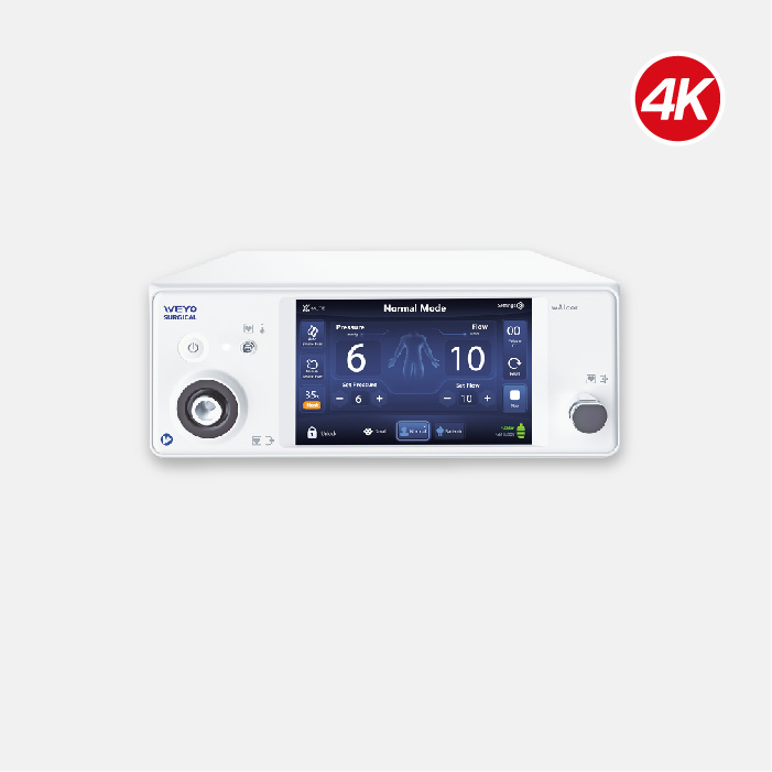
Then there’s the monitor, right? If the feed coming out of the processor is Full HD, you need a monitor that can actually display it correctly. A lot of hospitals skip this and use old office monitors, which are dim or have bad color accuracy. Our systems pair with industrial-grade Full HD monitors that are calibrated to show every color exactly how it is, and they’re bright enough to work in the OR’s operating lights—no squinting to see what’s on screen. Some of our newer setups even have 4K monitors, but most arthroscopies don’t need that extra resolution, and 1080p is more than enough for the tiny tools and 1mm structures surgeons work with. Wait, let’s not forget about the video recording and archiving part, either. Most hospitals need to keep surgery footage for patient records, malpractice, or training. Our systems record directly to a secure drive, no extra equipment needed, and you can label procedures in 10 seconds flat—no more filling out messy paper logs after a long day.
Now, let’s talk about why Full HD is non-negotiable for arthroscopies specifically, not just any endoscopic surgery. Arthroscopic joints are super confined—like, a knee joint is only big enough to fit your thumb and index finger side by side, right? So the scope is only a few millimeters thick, and the camera tip is even smaller. If the image is grainy, the surgeon can’t see the edge of a tear, so they might remove too much healthy meniscus, which leads to arthritis later. Or they might not see a small loose bone fragment in the joint, which would keep the patient in pain after surgery. I had a customer who was using a competitor’s cheaper Full HD system, and he had a case where he missed a tiny tear in the lateral meniscus because the image was washed out. He switched to ours, and he said he’s had way fewer post-op follow-ups for “unseen damage.” That’s the real impact of this tech—its not just about crisp video, it’s about better patient outcomes.
Wait, there’s a common myth I hear all the time: “Oh, DSLR cameras have better HD, why can’t arthroscopic systems just use that?” Let me explain why that’s not the case. DSLRs are built to take photos of large, well-lit scenes, with room to adjust. Arthroscopic scopes have to fit through a tiny tube, work in a dark, sealed space, with light that can’t be too bright (you don’t want to burn tissue, right? That’s a huge safety thing). The sensors in arthroscopic cameras are way smaller than even the smallest DSLR sensor, so they have to be optimized to capture clear, bright, true-color video in those tight conditions. Our sensors are custom-built for exactly that—they don’t work for Instagram, but they work perfectly for surgery. That’s a big difference between a $1000 off-the-shelf camera and a $10,000 medical-grade scope system.
Another thing that’s easy to overlook: ergonomics for the surgeon. If the monitor is grainy or laggy, the surgeon has to lean way in to see it, which causes neck and back pain over time. I’ve talked to senior ortho surgeons who’ve been doing scope work for 20 years, and their main complaint with old systems is that they end the day with a stiff neck. Our Full HD monitors are positioned at eye level, and the feed is so smooth they don’t have to hunch over as much. That might sound trivial, but a surgeon with chronic pain can’t operate as well, and they can’t work as long. It’s a big deal for retention and consistent quality of care.
Let’s also touch on how the system integrates with other OR tech, because that’s a huge selling point for hospitals that are updating their whole surgical suite. Our Full HD endoscopic systems work with robotic scope holders, so the surgeon can control the scope with a foot pedal or voice command instead of an assistant. That means less handoffs, less chance of the scope slipping, and more control during delicate procedures. For example, when repairing a rotator cuff, the surgeon needs to keep the scope steady while they tie sutures, and a robotic holder paired with our clear feed makes that so much easier. A lot of older systems don’t play nice with robotic tools, but we designed ours to integrate with all the main OR automation brands, so hospitals don’t have to replace every piece of gear to upgrade their endoscopes.
Now, I know you’re probably wondering: is Full HD worth the cost? Let’s be real, medical equipment isn’t cheap, but the ROI is there. Faster surgery times because surgeons don’t have to search for damage, fewer post-op complications, less time in the OR, and shorter patient stays. I had a hospital in Ohio that switched from a 720p system to our Full HD setup, and their average arthroscopic surgery time dropped by 12 minutes. Multiply that by 500 surgeries a year, and that’s 100 hours of OR time saved—time they can use for other patients, which translates to more revenue for the hospital and better access for people waiting for surgery. That’s not a sales pitch, that’s math.
If you’re reading this, you’re probably a hospital administrator, a surgery manager, or an orthopedic surgeon looking to upgrade your scope systems. Maybe you’re tired of grainy images, laggy feeds, and scrub techs fumbling with light settings mid-op. Or maybe you’re a rep looking to update the systems at your clients’ facilities. Either way, our Full HD endoscopic systems are built for real OR use, not just trade shows. We’ve tested them in over 200 hospitals across North America, and the feedback has been consistent: clearer images, faster surgeries, happier surgeons and patients.
If you’re ready to learn more about how our systems can work for your clinic or hospital, just reach out to our team for a no-obligation demo and consultation. We can send a rep to your facility to show you how the system works, let your surgeons test it on phantom models, and answer any questions you have about integration, training, or pricing. No pressure, no sales jargon, just straight answers about how our Full HD tech can make a real difference in your arthroscopic surgeries.
Endoscopy Related Equipments References
- American Academy of Orthopaedic Surgeons. Arthroscopic Surgery: What to Expect. 2022.
- Lee SJ, et al. The Impact of High-Definition Imaging on Arthroscopic Diagnostic Accuracy. Journal of Arthroscopy and Related Surgery, vol. 36, no. 8, 2020, pp. 2145-2151.
- World Health Organization. Medical Device Regulation for Surgical Endoscopy Systems. 2021.
- Smith AB, et al. Ergonomic Improvements in Full HD Endoscopic Systems for Orthopedic Surgeons. Surgical Endoscopy, vol. 35, no. 12, 2021, pp. 6789-6795.
Jiangsu Bonss Medical Technology Co., Ltd.
As one of the most professional full hd endoscopic system manufacturers and suppliers in China, we’re featured by quality products and good price. Please rest assured to buy bulk full hd endoscopic system for sale here from our factory. Customized orders are welcome.
Address: Building #7,No.898,China Medical City Avenue, Haling District, Taizhou City.Jiangsu P.R. China
E-mail: inquiry@plasma-surgical.com
WebSite: https://www.bonssmedical.com/
